Biology Reference
In-Depth Information
DsRed
DsRed2
Fluorescent
Timer
DsRed
N42H
AG4
500
600
700
500
600
700 500
600
700
λ
/nm
λ
/nm
λ
/nm
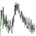
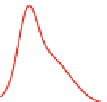
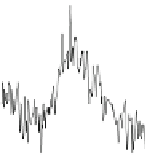
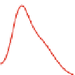
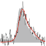
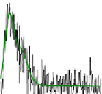
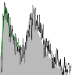
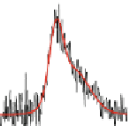
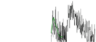
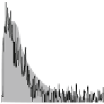
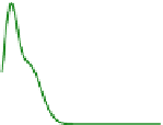
Fig. 9 Single tetramer spectra of DsRed and the analyzed variants. For all proteins emission
spectra showing solely green emission (
first column
), mixed emission (
second column
), or solely
red emission (
third column
) were found. The emission from the red-emitting chromophore was
systematically shifted to the red, consistent with the fast formation of a super-red form. The
frequencies of occurrence of the respective spectra were consistent with the bulk spectra, displayed
in grey behind the predominantly observed spectrum (From [
88
], Copyright Wiley-VCH Verlag
GmbH & Co. KGaA. Reproduced with permission)
Indeed, all variants for which the super-red form was found contain a glutamate
residue at position 215; thus, the universal observation of the super-red form
is consistent with this proposed mechanism. However, the super-red form was


Search WWH ::

Custom Search