what-when-how
In Depth Tutorials and Information
also reported increased fluoride in the oim
/
oim animals
in different backgrounds. This increase in fluoride con-
tent could be explained based on the smaller size, and
hence greater surface area, of the bone mineral crystals.
fraction), trabecular number, and cortical and trabecular
density after transplantation of hematopoietic stem cells
into the oim
/
oim mice
68
and showing that treating this
same mouse model with either a bisphosphonate or an
inhibitor of RANK ligand caused significant increases in
trabecular number, decreased trabecular thickness and
separation.
66
Neither micro-CT of biopsies from humans
with OI nor high-resolution peripheral quantitative
CT studies of human OI bone have yet been reported,
although with the introduction of high-resolution CT
69
it seems likely that this type of data should soon be
available.
MICRO-COMPUTED TOMOGRAPHY
Three-dimensional images of bone density obtained
by micro-computed tomography (micro-CT) and
two-dimensional representations of such images pro-
vide information on the geometry and microarchi-
tecture including connectivity of bone (
Figure 4.6
).
Measurements can be made
ex vivo
on tissues in water or
alcohol, or
in vivo
on small animal bones. These analyses,
which are based on images thresholded to remove the
presence of solvent or even marrow, provide information
on the amount of bone (cortical or cancellous) present,
the presence of fractures, the thickness and area of the
bone examined, the number and size of trabeculae, and
their connectivity and shape.
65,66
Micro-CT was used to describe the phenotype of a
wide variety of mouse models of OI including the oim
/
oim,
66
the BrtlIV,
45
the mouse model of a form of OI
found in an Amish population
47
and fro
/
fro mice.
54
OI
bone from a novel animal model (knockout of the osteo-
potentia (OPT) protein) was first described based on
micro-CT. In the OPT study, not only did the micro-CT
details agree with both histology and standard X-rays,
but micro-fractures not visible in the plain films were
apparent using micro-CT.
67
In general, in mouse models
of OI, two- and three-dimensional micro-CT studies have
documented an increase in mineral density per unit area,
a thinning of trabeculae and cortices, and a reduced cor-
tical bone width.
Micro-CT has been useful in assessing the effects of
agents used to treat OI, demonstrating, for example, sig-
nificant increases in the amount of bone (bone volume
Recent Biochemical and Genetic Analysis Related
to Mineralization Processes
What is now considered classical biochemistry, along
with genetic studies reviewed elsewhere, identified col-
lagen and related gene defects associated with collagen
production in patients with different forms of OI.
70-73
But
with the exception of the last reference,
73
the question of
which, if any, of the other proteins associated with min-
eralization have altered expressions has been addressed
to a much lesser extent.
Compositional analysis of cortical bone from human
biopsies of 26 OI patients (types I, II, III and IV) and
seven age-matched controls demonstrated reduced lev-
els of osteonectin, increased levels of osteocalcin and
alpha-2HS-glycoprotein in all OI patients, and elevated
bone sialoprotein with the highest levels only in type
IV OI.
74
Osteonectin, which is co-expressed with colla-
gen, binds to collagen and regulates collagen-fibril size,
75
and most likely is reduced in parallel with the reduction
in collagen expression. Osteocalcin regulates bone turn-
over, among other activities,
76
and may be elevated due
to its binding to the smaller mineral crystals which have
a larger surface
/
volume ratio. Alpha-2-HS glycoprotein
(A)
(B)
100
µ
m
100
µ
m
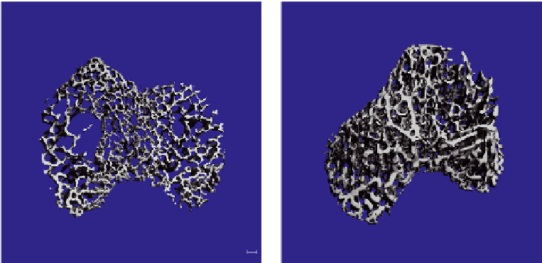
FIGURE 4.6
Micro-CT three-dimensional images of (A) Amish model of OI (OOA
/
+) and (B) wild-type (+
/
+) trabecular bone at 2 months.
Notice the increased porosity in the OAA
/
+ mice.