what-when-how
In Depth Tutorials and Information
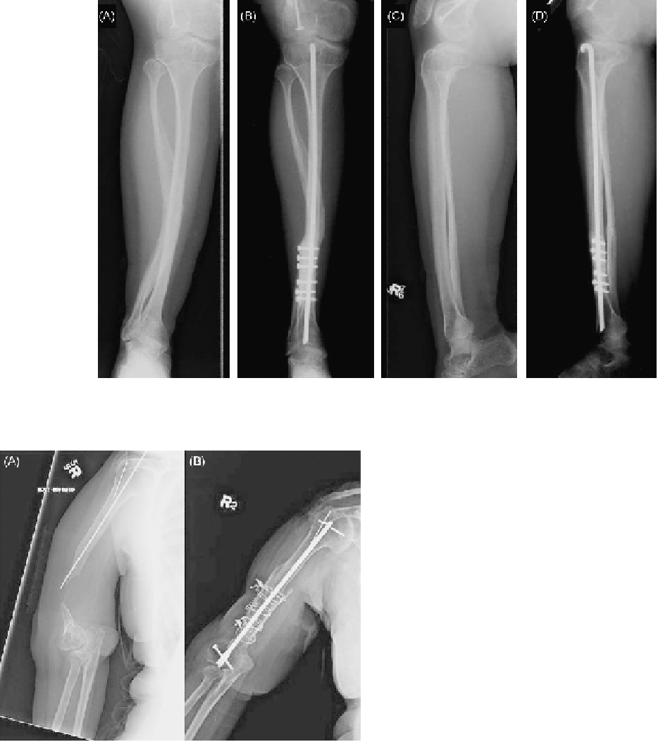
FIGURE 48.1
Correction of valgus deformity in tibia with osteotomy, intramedullary rod and femoral allograft: (A) preoperative anterior
posterior view; (B) postoperative anterior posterior view; (C) preoperative lateral view; (D) postoperative lateral view.
being tibial or fibular osteotomies. In three additional
procedures, pre-existing fractures were used to realign
bones rather than creating new osteotomies. The osteot-
omies were then stabilized by the addition of bridging
allografts, implants or both. In many cases, other types
of graft material, including demineralized bone matrix
and cancellous cubes, were added to the osteotomy
sites to promote osteogenesis and healing.
Allograft Struts
Allografts were used to stabilize osteotomies or
non-unions and promote osteogenesis.
Table 48.2
sum-
marizes the strut allografts implanted by source bone
and site of implantation. Fourteen strut allografts were
implanted in ten patients during 13 procedures.
In many cases, a sandwich-type allograft was
implanted, in which two pieces of allograft were shaped
to it the native bone and held in compression using
additional implants (
Figure 48.2B
). Allograft in the form
of a “sandwich” provides both biologic and mechani-
cal benefits, especially in diaphyseal fixation. As the
two halves of the sandwich are compressed, they pro-
vide a high degree of frictional stabilization against
rotation because of the large contact area. They have
been observed to incorporate effectively, and periosteal
bridging between the allograft and the diaphysis may
be observed after 2-3 months in many patients. The
allograft should span at least 3 cm on each side of the
osteotomy or defect. Each half should cover at least 25%
FIGURE 48.2
Stabilization of chronic non-union in humerus with
radial allograft and intramedullary rod: (A) preoperative image; (B)
postoperative image.
SURGICAL TECHNIQUES
Osteotomies
Osteotomies were performed on most patients
undergoing limb reconstruction, with correction of a
limb deformity or joint misalignment as the primary
indication (
Figure 48.1B
). The osteotomies are sum-
marized by location in
Table 48.1
, with over one-half