what-when-how
In Depth Tutorials and Information
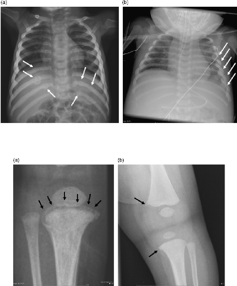
FIGURE 42.1
(a) Healing bilateral posterior rib fractures in a 9-month-old infant. (b) Healing LT lateral rib fractures found in a 44-day-old
infant with severe intracranial injury (see
Figure 42.5
).
FIGURE 42.2
(a) “Bucket-handle” appearance of CML of the RT proximal tibia in a 3-month-old infant with intracranial hemorrhage.
(b) “Corner appearance” of CML LT distal femur and proximal tibia in a 56-day-old with “leg injury.”
fracture. CMLs should raise concerns even if the patient
has been diagnosed with OI.
Long bone shaft fractures are frequently seen in
NAT.
7,11,16
The fracture may be acute and may be the
presenting symptom, it may be healing with subperios-
teal elevation, or it may be both acute and in the process
of healing (
Figure 42.3a,b
). The type of fracture seen
radiographically suggests a certain underlying mecha-
nism of injury, therefore a detailed history obtained by
the child abuse pediatrician or other medical provider
is of the utmost importance to ascertain the plausibility
of the fracture sustained. A spiral fracture usually indi-
cates a torsion mechanism, while a transverse fracture
usually indicates that the bone has hit an object, such
as during a fall, or that an object has struck the bone.
However, spiral fractures may occur in OI depending
on the type of injury. A buckle component can indicate
a degree of impaction. Oblique fractures are often due
to a combination of forces. However, more important
than the type of fracture is the developmental stage of
the child and ability/inability of the child to incur such
a fracture. For example, a 3-month-old who sustains a
transverse fracture of the femur is more cause for con-
cern, compared to a 2-year-old who sustains a spiral
fracture of the femur after tripping while running.
Virtually any bone in the body can be fractured
including the spine, small bones of the hand and the
feet, and the axial skeleton. Fracture of the acromion,