what-when-how
In Depth Tutorials and Information
softening of the occipital bone
10
and repeated craniover-
tebral junction microfractures that heal in an abnormal
fashion.
11
Patients with BI can present with a myriad of features
that arise as a consequence of brain stem and cerebellum
compression, disturbance of cerebrospinal fluid dynam-
ics, mechanical stretching of cranial nerves and even from
vertebrobasilar blood low obstruction.
3,11
These features
can be progressive, and can lead to rapid neurological
deterioration, respiratory arrest or even sudden death.
16,17
CLINICAL SYMPTOMS
The majority of OI patients with BI present with
severe physical disability and advanced symptomatic
BI. Ibrahim et al., in a retrospective review of outcomes
in 20 patients, reported that 40% had sleep apnea, 60%
were regular wheelchair users, 80% had muscular weak-
ness with pyramidal signs, 45% had dysphagia, 20% had
a diminished gag reflex and 25% were found to also suf-
fer from vertigo.
3
Lower cranial nerve abnormalities
involving cranial nerves VIII, IX and X have also been
described in 70% of OI patients with BI.
2
At presenta-
tion, 15 out of 20 patients scored 70% or lower on the
Karnofsky Performance Scale (KPS), whereas the remain-
ing five patients scored 80% or higher. According to the
authors, the three most common clinical symptoms were
nystagmus, ataxia and headaches. Severe kyphoscoliosis
was present in almost one-half of the patients. Moreover,
the Ranawat classification for neurological deficits and for
pain can be used to evaluate myelopathy and pain.
18
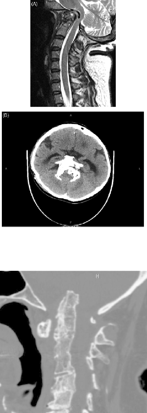
FIGURE 36.1
(A) Sagittal MRI image demonstrating basilar invagi-
nation in OI with brainstem compression. (B) Axial CT of the head dem-
onstrating high-riding osseous structures due to basilar invagination.
(Reproduced with permission). Copyright © 2014 Jean-Paul Wolinsky.
DIAGNOSIS
Once there is ample clinical suspicion of BI in patients
with OI, several radiological modalities are utilized to
confirm the diagnosis and aid in preoperative planning.
In OI, lateral skull radiographs have been recommended
as a screening method at individually adjusted intervals.
A baseline cephalometric study has been suggested for
all OI patients before school age.
19
Plain radiographs
and computed tomography (CT) images delineate the
bony anatomy and alignment, while magnetic reso-
nance (MR) imaging detects neural element compres-
sion, presence of syrinx and soft tissue abnormalities.
Chamberlain's line, Wackenheim's clivus baseline, the
clivus-canal angle, the basal angle as well as the ante-
rior atlanto-dental interval can be used in assessment of
atlanto-axial instability.
20
McGregor introduced a modi-
fication of Chamberlain's line, namely “the line drawn
from the upper surface of the posterior edge of the hard
palate to the most caudal point of the occipital curve in
the true lateral X-ray.”
21
Three measures on lateral skull
FIGURE 36.2
Cranial settling due to C1-C2 instability.
(Reproduced
with permission). Copyright © 2014 Jean-Paul Wolinsky.