what-when-how
In Depth Tutorials and Information
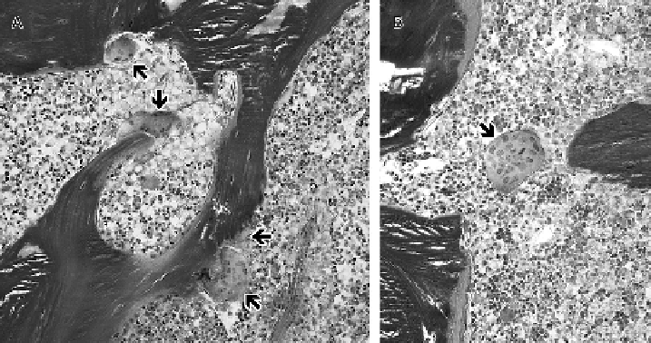
FIGURE 25.3
Iliac bone specimen from a 17-year-old adolescent with OI type IV after 2.9 years of pamidronate treatment. Osteoclasts are
marked by arrows. The magnification bar corresponds to 100 μm. (A) Both osteoclasts with bloated appearance and osteoclasts with normal size
are visible. (B) Large osteoclast, which appears to be detached from the bone surface.
(From: Cheung MS, Glorieux FH, Rauch F. Large osteoclasts in
pediatric osteogenesis imperfecta patients receiving intravenous pamidronate. J Bone Miner Res 2009;
24
: 669-74.)
increased by 87% and 38%, respectively, between base-
line and the first time point during treatment. Thereafter,
cortical width did not change significantly, but there was
a trend towards higher cancellous bone volume. Average
bone formation rate on trabecular surfaces decreased
by 70% after pamidronate treatment was initiated, and
showed a trend towards a further decline in the second
part of the study interval. These results indicate that the
gains that can be achieved with pamidronate treatment
appear to be largely realized in the first 2 to 4 years.
Little is known about the bone tissue-level effects of
bisphosphonate compounds other than pamidronate.
One study examined the efficacy and safety of oral rise-
dronate in the treatment of pediatric patients with mild
OI.
41
Iliac bone biopsies were performed at the end of
the 2-year study period. Histomorphometric analysis of
these transiliac bone biopsies did not show a significant
treatment difference in cortical width, trabecular bone
volume or parameters of bone turnover. These results
suggest that the skeletal effects of oral risedronate are
weaker than those that are commonly observed with
intravenous pamidronate treatment.
The effect of bisphosphonate treatments in adults
with OI has been studied in one case series.
42
This
revealed an increase in both trabecular bone volume
and in cortical thickness. Thus, the structural effect in
adults was similar to those in children, even though the
magnitude of the effect seems to be smaller in adults.
appearance of transverse lines in the metaphyses of long
bones. These lines were examined in the case of a child
with OI type VII, where the iliac bone biopsy had inad-
vertently included part of the iliac growth plate and the
adjacent metaphysis.
43
It was seen that these metaphy-
seal lines corresponded to horizontal trabeculae that
were undergoing active remodeling, rather than “frozen
growth plate cartilage” as had been hypothesized before.
Changes in the appearance of osteoclasts have previ-
ously been noted in children receiving pamidronate and
have been interpreted as signs of toxicity.
35,44
A study
analyzed osteoclast parameters in paired iliac bone speci-
mens before and after 2 to 4 years of cyclical intravenous
pamidronate therapy in 44 pediatric OI patients and
found that intravenous pamidronate of young OI patients
leads to an increase in osteoclast size (
Figure 25.3
).
45
However, the presence of large osteoclasts was not associ-
ated with detectable adverse clinical effects.
References
Adverse Effects of Bisphosphonate Treatment on
the Tissue Level
One of the radiological features of intravenous
bisphosphonate treatment in growing children is the