what-when-how
In Depth Tutorials and Information
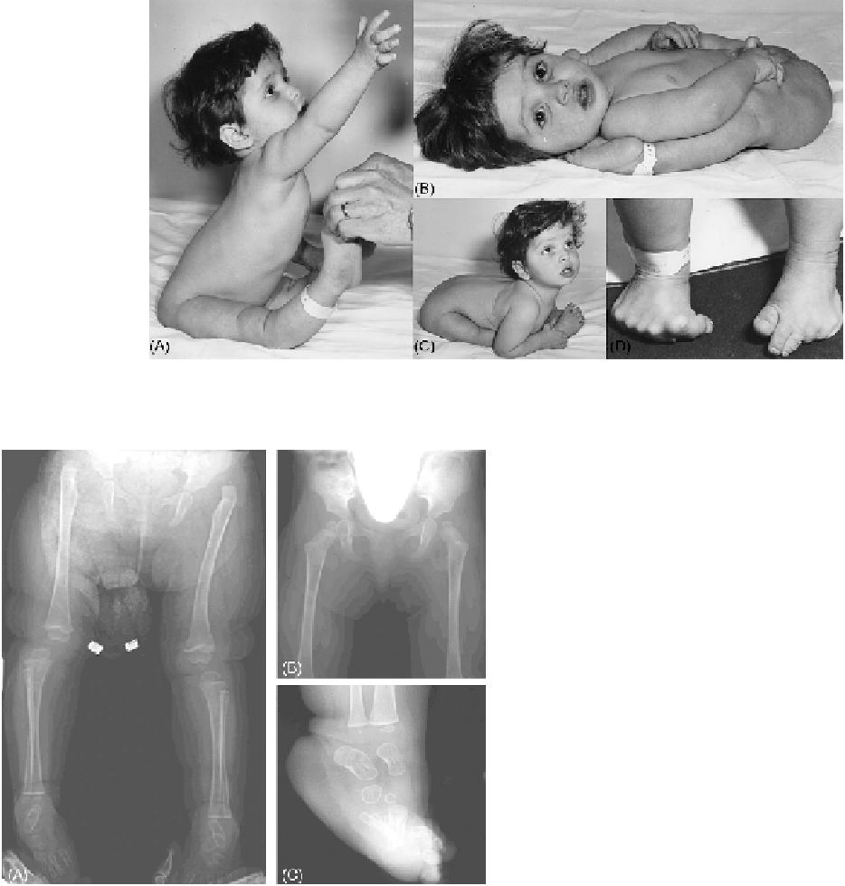
FIGURE 23.1
A 12-month-old girl with EDS VIIB (Case 18) before correction of the hip dislocation. (A) Subluxations of both knees, moder-
ate thoraco-lumbar kyphosis. (B) Congenital hip luxations. (C, D) Marked hyperlaxity of the small joints.
(Reprinted by permission from
2
.)
Studies by Wirtz et al.
23
showed that in EDS VIIB
(Case 18), the α1(I) N-propeptide, although cleaved
from the α1(I) chain by the N-proteinase or (less likely)
by non-specific proteases, is retained by non-covalent
association with the mutant pNα2(I) chain in native
mutant collagen molecules, both
in vivo
and
in vitro
;
the α1(I) N-propeptide was readily demonstrable by
Western blotting of skin extracts, and rotary shadow-
ing of pepsin-treated collagen produced in culture dis-
closed kinked molecules which were longer than their
normal counterparts (
Figure 23.4E
). These data suggest
that the retention of a partially cleaved, but essentially
intact, N-propeptide in mutant collagen may play a crit-
ical role in the pathogenesis of this disease.
Morphology of the Collagen Fibrils
The skin collagen fibrils of EDS VII patients have a
smaller and more variable diameter than normal. They
are irregular in outline, and also appear to be more
loosely and randomly organized into fibrils resembling
a loosely wound rope (
Figure 23.5
). These changes are
more pronounced in the patients affected with EDS
VIIA (
Figure 23.5A
) compared to those observed in EDS
VIIB patients (
Figure 23.5B
), but less severe than those
present in EDS VIIC (
Figure 23.5C
; see “Genotype-
Phenotype Correlation,” below).
FIGURE 23.2
Radiographs before and after open reduction of the
hip dislocation in a patient with EDS VIIA (Case 37). (A) Radiograph
of the pelvis from the patient at 12 months of age shows bilateral
luxation of the hips, and anterolateral subluxation of the knees due
to severe ligamentous instability. (B) Radiograph of the pelvis from
the patient at 23 months of age, 6 months after open reduction, shows
successful correction of bilateral dislocation of the hips. (C) A radio-
graph of the right foot shows the disorganization of the metatarsal
bones. Please note the striking osteopenia which radiologically seems
more pronounced than in EDS VIIB (compare with
Figure 23.3
).
(Reprinted by permission from
35
.)
Genetics
EDS VIIA and VIIB are inherited as autosomal dom-
inant traits, and some of the sporadic cases have been
shown to be heterozygous for a new mutation (
Table
23.1
). Mosaicism in the mother of Case 13 is likely the
cause of her milder phenotype as opposed to her four
respectively. The pNα2(I)-like chains migrate between
the α1(I) and the α2(I) chains of collagen I (
Figure 23.4F
),
whereas the pNα1(I)-like chains migrate just above the
normal α1(I) chains, and thus their band is more difficult
to detect.