Plus-Strand RNA Viruses
the relationships of these viruses to the plant viruses and what
INTRODUCTION
this means in terms of virus evolution.
The plus-strand RNA [(+)RNA] viruses comprise a very
large group of viruses belonging to many families. Among
FAMILY PICORNAVIRIDAE
these are viruses that cause epidemic disease in humans,
including encephalitis, hepatitis, polyarthritis, yellow fever,
The picornaviruses are so named because they are small
dengue fever, poliomyelitis, and the common cold. The
(pico = small), RNA-containing viruses. Nine genera of
number of cases of human disease caused by these viruses
picornaviruses, five of which contain human pathogens, are
each year is enormous. As examples, dengue viruses infect an
currently recognized (Table 3.2), and more will probably
estimated 50 to 100 million people each year; most humans
be recognized as further studies of the known viruses occur
suffer at least one rhinovirus-induced cold each year, with the
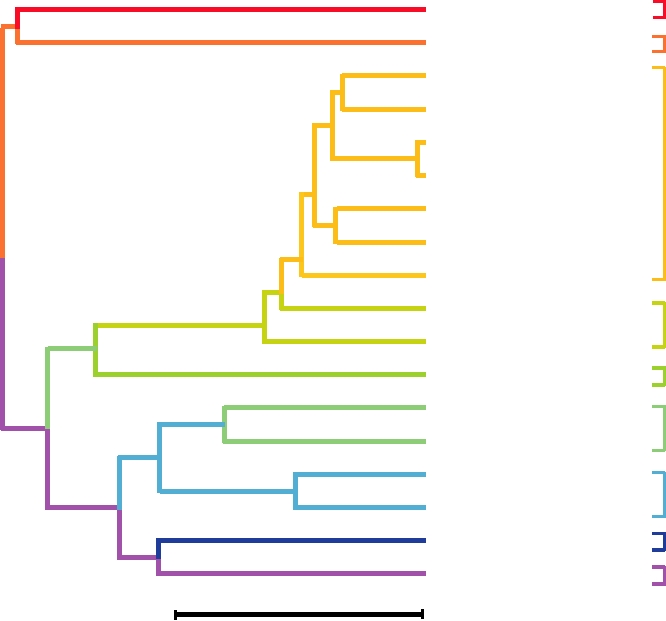
and as new viruses are described. A dendrogram that illus-
cases therefore numbering in the billions; and most humans
trates the relationship of the nine genera to one another,
during their lifetime will suffer several episodes of gastro-
as well as the relationships of a number of viruses within
enteritis caused by astroviruses or caliciviruses. In terms of
the various genera, is shown in Fig. 3.1. This dendrogram
frequency and severity of illness, the (+)RNA viruses con-
makes clear that all picornaviruses are closely related.
tain many serious human pathogens, and we will begin our
They share significant nucleotide and amino acid sequence
description of viruses with this group.
identity and form a well-defined taxon. The dendrogram
The human (+)RNA viruses belong to seven families
also illustrates the rationale for grouping these viruses into
(Table 3.1). These seven families also contain numerous
nine genera.
viruses that infect other vertebrates, of which many are
As described in Chapter 2, the structures of several picor-
important pathogens of domestic animals. Large numbers of
naviruses have been solved to atomic resolution by X-ray
(+)RNA viruses that infect plants are also known; in fact, most
crystallography. The picornavirus virion is composed of 60
plant viruses contain (+)RNA genomes. The plant viruses,
copies of each of four different proteins (called VP14) that
however, belong to different families and are currently classi-
form an icosahedral shell having T=3 symmetry (or pseudo-
fied by the International Committee on Taxonomy of Viruses
T=3) and a diameter of approximately 30 nm (see Figs. 2.1,
(ICTV) into nine families plus many unassigned genera.
2.5, 2.7, and 2.8).
Because of their importance as disease agents of domestic
crops, much is known about these viruses. Other families of
(+)RNA viruses include two families of bacterial viruses,
Organization and Expression of the Genome
one of fungal viruses, and four families of insect viruses (the
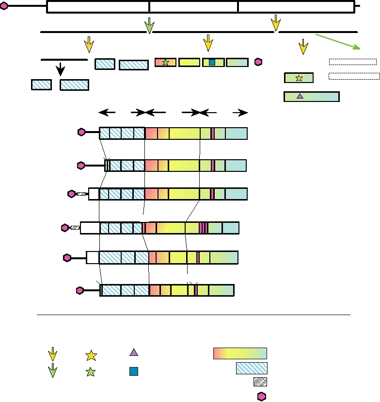
The structure of the genome of poliovirus and comparison
nodaviruses, in particular, have been intensively studied).
of it with the genomes of the other genera are shown in Fig.
Thus, the (+)RNA viruses have evolved into many distinctly
3.2. The picornaviral genome is a single RNA molecule of
different families and must have arisen long ago. In this chap-
about 7.5 kb. It contains one open reading frame (ORF) and
ter, the seven families of viruses that include human viruses
is translated into one long polyprotein. This polyprotein is
as members are considered, followed by a brief discussion of
TABLE 3.1
Families of Plus-Strand RNA Viruses That Contain Human Pathogens
Family
Size of genome (nucleotides)
Other vertebrate hosts
Representative human pathogens
∼7500
Picornaviridae
Cattle, primates, mice
Poliovirus
Human rhinovirus
Hepatitis A
∼7500
Caliciviridae
Rabbits, swine, cats
Norwalk
Hepeviridae
7200
Primates, swine
Hepatitis E
Astroviridae
68007900
Cattle, ducks, sheep, swine
Human astrovirus
∼11,600
Togaviridae
Mammals, birds, horses
Semliki Forest, Ross River, WEE, VEE, EEE,
Mayaro, rubella
Flaviviridae
950012,500
Swine, cattle, primates, birds
Dengue, yellow fever, JE, MVE, TBE, WNV,
hepatitis C
Coronaviridae
20,00030,000
Mice, birds, swine, cattle, bats
SARS coronavirus
Virus name abbreviations: WEE, VEE, EEE, Western, Venezuelan, Eastern equine encephalitis viruses; JE, Japanese encephalitis virus; MVE, Murray Valley
encephalitis virus; TBE, tick-borne encephalitis virus; WNV, West Nile virus; SARS, severe acute respiratory syndrome.
TABLE 3.2 Picornaviridae
Virus name abbreviationa
Genus/members
Usual host(s)
Transmission
Disease
World distribution
Enterovirus
Human enterovirus A
HEV-A
Humans
Oralfecal, contact
See Table 3.5
Worldwide
Human enterovirus B
HEV-B
Humans
Oralfecal, contact
See Table 3.5
Worldwide
Human enterovirus C
HEV-C
Humans
Oralfecal, contact
See Table 3.5
Worldwide
Human enterovirus D
HEV-D
Humans
Oralfecal, contact
See Table 3.5
Worldwide
Poliovirus (Types 1,
PV
Humans
Oralfecal, contact
See Table 3.5
Originally worldwide,
2, and 3)
extirpated in Americas
Bovine enterovirus, porcine enteroviruses A and B; Unassigned enteroviruses of humans and monkeys
Rhinovirus
Human rhinoviruses
HRV-A, HRV-B
Humans
Aerosols, contact
Common cold
Worldwide
(>100 serotypes)
Cardiovirus
Encephalomyocarditis
EMCV
Mice
Oralfecal, contact
Encephalitis,
Worldwide
Theilovirus
myocarditis
Aphthovirus
Foot and mouth disease
FMDV
Cattle, swine
Oralfecal, contact
Lesions on
Worldwide (except
mouth and feet
United States,
Australia)
Equine rhinitis A
ERAV
Horses
Worldwide
Hepatovirus
Hepatitis A
HAV
Humans
Oralfecal
Hepatitis
Endemic worldwide
Parechovirus
Human parechovirus
HPeV
Humans
Oralfecal
Gastroenteritis
Worldwide
Erbovirus
Equine rhinitis B
ERBV
Horses
?
?
?
Kobuvirus
Aichi
AiV
Humans
Oralfecal
Gastroenteritis
Isolated in Japan (oysters)
Teschovirus
Porcine teschoviruses
PTV-1
Swine
Oralfecal
Paralysis, porcine
Britain, central and
(10 species recognized)
encephalomyelitis Eastern Europe
a
Standard abbreviations are given for either the virus listed (such as poliovirus) or for the type member of the genus.
Hepatovirus
Hepatitis A
Parechovirus
Human parechovirus 1
Human enterovirus B
Human enterovirus D
Human enterovirus C
Enterovirus
Poliovirus type 1
Bovine enterovirus 1
Porcine enterovirus 1
Human enterovirus A
Human rhinovirus 14
Rhinovirus
Human rhinovirus 2
Kobuvirus
Aichi
Equine rhinitis A
Aphthovirus
Foot and mouth disease
Encephalomyocarditis
Cardiovirus
Theilovirus
Erbovirus
Equine rhinitis B
Teschovirus
Porcine teschovirus
Evolutionary Distance
0.5
FIGURE 3.1 Relationships between 18 representative picornaviruses. The viruses shown have been classified into the 9
recognized genera. This tree was generated from the amino acid sequences of the 3Dpol proteins. Adapted from Yamashita et
al. (2003), and updated with the taxonomy found in Fauquet et al. (2005). Evolutionary distance, calculated by the UPGMA
(unweighted pair group method with averages) method, is the number of residue substitutions that have occurred between
two sequences since their divergence from a common ancestor and is defined as D = number of base mismatches/total
alignment length in nucleotides.
cleaved by one or more virus-encoded proteinases to form
of RNA synthesis. VPg is normally removed from RNA that
more than 25 different polypeptides, including processing
serves as mRNA by a cellular enzyme, but its removal is not
required for its translation. The 3′ end of the RNA is poly-
intermediates (not all of which are shown in the figure) as well
adenylated. As described in Chapter 1, the 5′ nontranslated
as final cleavage products. The ORF in the genome contains
three regions, called P1 (the 5′ region), P2 (the middle region),
region of a picornaviral RNA possesses an IRES (internal
and P3 (the 3′ region). Region 1 encodes the structural proteins
ribosome entry site) and the RNA is translated by a cap-inde-
and regions 2 and 3 encode proteins required for RNA replica-
pendent mechanism. The translation of picornaviral RNA is
tion. The genome organization of all picornaviruses is similar,
greatly favored in the infected cell because picornaviruses
but each genus differs in important details. For example, the
interfere with host cell macromolecular synthesis and, in
aphthoviruses and the cardioviruses have a poly(C) tract near
particular, interfere with host protein synthesis. Infection
the 5′ end of the RNA that is important for virus replication.
with entero-, rhino-, and aphthoviruses leads to proteolytic
These two genera also have a leader polypeptide that precedes
cleavage of a cellular protein called eIF4G that is a compo-
the structural protein region. The aphthovirus leader peptide is
nent of the cap-binding complex. Cleavage of this protein by
2Apro of entero- and rhinoviruses or by the leader protease
a papain-like protease that cleaves itself from the polyprotein
and has a role in the shutoff of cellular protein synthesis. The
of aphthoviruses results in inhibition of the translation of
function of the cardiovirus leader is not known. Hepatitis A
RNAs that require the cap-binding protein complex, that is,
virus, Aichi virus, and echovirus 22, representatives of three
capped host cell mRNAs. The cardioviruses, which are also
other genera, also have leaders.
cap independent, interfere with translation of host mRNAs
The picornaviral genome has a small protein, VPg, cova-
in a different way, by interfering with phosphorylation of
lently bound to the 5′ end, which is the primer for initiation
cap-binding protein. Poliovirus also interferes with host
protein synthesis by cleavage of poly(A)-binding protein by
illustrated for poliovirus in Fig. 3.2. Some cleavages occur in
the viral 3Cpro, but the mechanism by which this interference
cis and some in trans. The crystal structures of 3Cpro of polio-
operates on cellular protein synthesis and not viral protein
virus and of a rhinovirus have been solved to atomic resolu-
synthesis, since poliovirus RNA is also polyadenylated, is
tion, and their core structure resembles that of chymotrypsin
not yet clear. In addition, 3Cpro of apthoviruses cleaves ini-
(Fig. 1.19A). The catalytic center has the same geometry as
that of chymotrypsin, but in 3Cpro the catalytic serine has
tiation factors eIF4A and eIF4GI, and it is thought that these
cleavages lead to a decrease in the level of viral protein syn-
been replaced by cysteine. Moreover, in many, but not all,
thesis later in infection, which facilitates packaging of the
picornaviruses the aspartic acid in the catalytic triad has been
replaced by glutamic acid. Thus, 3Cpro is related to cellular
viral RNA.
The viral 3Cpro and its precursor 3CDpro make multiple
serine proteases and may have originated by the capture of a
cleavages in the polyprotein translated from the genome, as
cellular serine protease during the evolution of the viruses.
A
2Apro
3Cpro
P1
P2A-3AB
P3CD
2Apro
pro
CDpro
3C
3C pro
2Apro
3
3B
2C
3A
VP3
VP1
2B
VP0
3C
?
3
3Cpro
D
VPg
VP4
VP2
3Dpol
P1
P2
P3
B
3B
3D
4
3
2
1 2A 2B
2C
3A 3C
Entero- and Rhinovirus
L 4
3B
2
3
1 2A 2B
2C
3A 3C 3D
Hepatovirus
3B
Cardiovirus
4 2
3
1
2A 2B
2C
3A 3C
3D
L
3B
2A
3B 3B
4
2
3
1
Aphthovirus
L
2B
2C
3A
3C 3D
2A
3B
VP0
3
1
L
2B
2C 3A 3C
3D
Kobuvirus
L
3A 3B
Parechovirus
1 2A 2B 2C
3D
3C
VP0
3
Viral Proteases
Replicase Motifs
Coding Domains
Cleavages Enzymes
3Cpro
Nonstructural proteins
Polymerase (GDD)
3CDpro
Structural proteins
2Apro
Helicase
Poly(C) tract
Vpg
FIGURE 3.2 Genome organization of the Picornaviridae. (A) Genome organization of poliovirus showing the proteolytic
processing steps. Both the 3Cpro and 2Apro proteases are "serine-type proteases" with cysteine in the catalytic site. (B)
Comparative genome organizations of representatives of seven of the nine genera of Picornaviridae. The key to the different
shadings of coding domains and the symbols for various enzymatic motifs used in both (A) and (B) is given below. Adapted
from Murphy et al. (1995) p. 300 and Yamashita et al. (1998).
that this protease is unrelated to 2Apro of polioviruses, how-
Protease 3Cpro is present in all picornaviruses whereas pro-
ever. Thus, these two proteases in closely related
tease 2Apro is present in only a subset of picornaviruses. In
viruses have different origins, and the viruses have solved
poliovirus, 2Apro, like 3Cpro, is a serine protease in which the
the prob- lem of how to separate regions 1 and 2 in the
active site serine has been replaced by cysteine. 2Apro cata-
polyprotein in different ways. Furthermore, this finding
lyzes one essential cleavage in the polyprotein of poliovirus,
illustrates that recombination to introduce new functions into
that between P1 and P2. This cleavage occurs in cis. The pro-
viral genomes has been important in the evolution of these
teolytic activity of 2Apro is also required for other functions
viruses, a theme to which we will refer many times in this
topic. Still another solution to the problem of separating
during poliovirus replication, the nature of which have not
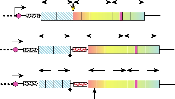
been established. An interesting experiment is illustrated in
regions 1 and 2 has been adopted by the cardio- and
Fig. 3.3 because it illustrates the power of molecular genetics
aphthoviruses. Protein 2A is not a protease in these viruses.
and the tricks that modern virologists can play with viruses.
Indeed, 2A is only 18 resi- dues long, and cleavage between
This experiment will serve as a prelude to the discussion of
P1 and P2 is catalyzed by 3Cpro. Another interesting feature
the uses of viruses as vectors in Chapter 11. A poliovirus was
of these viruses is that the cleavage at the 2A2B junction
constructed in which a stop codon was placed after the struc-
occurs spontaneously during translation, catalyzed by the
tural protein domain (region 1), so that 2Apro was not needed
specific amino acid sequence at the scissile bond. This
to remove P1 from the polyprotein precursor. The stop codon
cotranslational cleavage occurs only during translation on
was followed by an IRES and a new AUG start codon, so that
eukaryotic ribosomes, and it has been proposed that no
P2A and the rest of the genome could be translated from the
cleavage actually occurs, but that the 2A sequence
polycistronic RNA. This virus was viable. However, when
synthesis of the specific peptide bond at
prevents
the 2A proteinase was inactivated by changing the active site
the 2A2B junction.
In addition to these cleavages catalyzed by 2Apro, 3Cpro,
cysteine, the resulting virus was dead, showing that the prote-
olytic activity of P2A is required not only to separate regions
and the leader protease of aphthoviruses, VP0 is cleaved
1 and 2 of the polyprotein but also for other function(s).
during virion maturation to VP2 and VP4 in most, but not
Protein 2A of rhinoviruses is also a protease. The crystal
all, picornaviruses. Available evidence suggests that this
structure of protein 2A of human rhinovirus type 2 reveals
cleavage is not catalyzed by a protease.
3
5
Construct
2A Protease
Viable
P1
P2
P3
Activity
virus
T7
PV
3 NTR
3B
IRES
pT7PVXL2
++++
Large plaque
4
2
3
1 2A 2B
2C
3A 3C
3D
(wt)
T7
P2
P3
P1
EMCV
PV
3B
3 NTR
IRES
IRES
Not
Small plaque
4 2
3
1
2A 2B
2C
3A 3C 3D
pT7PVE2A
applicable
AUG
T7
P2
P3
P1
EMCV
PV
3B
3 NTR
IRES
IRES
pT7PVE2AX
None
Dead
4
2
3
1
2A 2B
2C
3A 3C
3D
AUG
C109A
FIGURE 3.3 Diagramatic illustration of constructs used to unravel the functions of protein 2A in poliovirus replication.
cDNA copies of the virus RNA can be manipulated by genetic engineering to insert IRES elements or make specific
mutations. RNA can be transcribed from the clones in vitro and used to infect cells, which is possible for plus-strand RNA
viruses because the first event after infection is translation of the genomic RNA. The wild-type construct pT7PVXL2 is
shown in the top line. The 2A proteolytic activity normally cleaves the bond between domains P1 and P2 of the translated
polyprotein. If this function is rendered nonessential, as in construct pT7PVE2A, by the insertion of a stop codon at the
C terminus of P1 (solid diamond), followed by a second IRES and an initiation AUG at the beginning of 2A, virus is still
produced, but forms small plaques. Thus separation of the structural region and the nonstructural region in this way results
in viable virus. However, if the proteolytic activity of 2A is inactivated by mutation of the catalytic cysteine to alanine as in
pT7PVE2AX, no virus is produced, demonstrating that the proteolytic activity of 2Apro is necessary for other functions in
addition to the P1/P2 cleavage. The pink hexagon is the VPg encoded in 3B and linked to the 5′ end of the RNA. Adapted
from Lu et al. (1995) and Molla et al. (1993).
RNA viruses. The 3A protein has hydrophobic sequences
Functions of the Picor navirus Proteins
that may be involved in this association. During replication, a
The cleavage product P1 consists of a polyprotein pre-
full-length complementary copy of the genomic RNA is pro-
cursor for the four structural proteins of the virus, VP14.
duced that serves as a template for the synthesis of genomic
P1 is first cleaved in trans to VP0, VP1, and VP3 by 3CDpro
RNA (illustrated schematically in Fig. 1.11A). This com-
(Fig. 3.2A). VP0 is later cleaved to VP2 and VP4 during
plementary RNA template has been variously called minus-
assembly of most picornaviruses.
strand RNA [abbreviated (-)RNA], antigenomic RNA, or
The cleavage products of P2 and P3 are required for
virion-complementary (vc) RNA. Much more (+)RNA than
RNA replication. 2Apro has been described. Protein 2B from
(-)RNA is produced, since (+)RNA is needed for translation
a Coxsackie virus has been shown to induce the influx of
and encapsidation into progeny as well as for replication,
extracellular Ca2+ and the release of Ca2+ from stores in the
whereas (-)RNA is needed only as a template for making
endoplasmic reticulum, and it is proposed that this protein
(+)RNA. It is probable that disproportionate amounts of (+)
induces lesions in cellular membranes that allow release of
and (-) strands are synthesized because the promoters in the
progeny virions. 2CATPase has been shown to be an ATPase,
viral RNA recognized by the viral replication machinery for
not a GTPase, and contains sequence motifs characteristics
(+) and (-)RNA synthesis (which might also be called ori-
of helicases. Many, but not all, RNA viruses encode heli-
gins of replication) differ in their strength, but other mecha-
cases to unwind duplex RNA during replication, and it is
nisms are known to be used in at least some RNA viruses.
assumed that 2CATPase performs such a function. The precur-
The RNA-dependent RNA polymerase 3Dpol is strictly
sor to 2B and to 2CATPase, a protein called 2BCATPase, has a dif-
primer dependent. In the presence of template, 3Dpol can uri-
ferent role in RNA replication. It is required for proliferation
dylate VPg on a specific tyrosine residue. This nucleotidyl
of membranous structures in poliovirus-infected cells that
peptide, VPgpU or VPgpUpU, then functions as a primer for
serve as sites for RNA replication.
the initiation of RNA synthesis. It is of interest that several
Region 3 encodes VPg, 3CDpro/3Cpro, and the viral RNA
viruses belonging to other families, such as hepatitis B virus
polymerase 3Dpol. Cleavages effected by 3Cpro are illustrated
(a virus that uses reverse transcription during the replica-
in Fig. 3.2. 3Cpro may also have a regulatory role in the virus
tion of its genome) and adenovirus (a DNA virus), have also
life cycle, because the cleavage intermediate 3CDpro, which
adopted the strategy of using a protein primer for initiation
is fairly long lived, has properties that differ from 3Cpro. One
of nucleic acid synthesis.
function of 3CDpro is to bind the viral RNA in conjunction
The nature and function of the promoters in the poliovirus
with 3AB, the precursor for VPg, or with a cellular protein,
genome that are involved in the initiation of RNA replica-
poly(C)-binding protein. Formation of a complex with the
tion are incompletely understood. One essential element has
viral RNA is essential for its replication, and differential
been called a cis-acting replication element, abbreviated cre,
cleavage of the 3C3D bond during the infection cycle may
or 3B-uridylation site, abbreviated bus. cre is a stem-loop
regulate replication. A strategy in which precursor proteins
structure that contains a motif in the loop, AAACA, that is
perform different functions than those performed by the final
conserved in all picornaviruses. This motif serves as a tem-
cleavage products, such as those illustrated by 2BCATPase and
plate for the uridylation of VPg described before, which is
3CDpro, allows the virus to optimize the coding capacity of
required for the initiation of RNA synthesis. The description
its small genome, because a given sequence is used for more
of this element as cis acting is a misnomer because the ele-
than one function.
ment can act in trans, and there is a pool of VPgpUpU within
poliovirus-infected cells that can be used to initiate RNA
synthesis. The cre element is found in different locations in
Replication of Picor naviruses
different picornaviruses. In poliovirus it is found in the cod-
The replication of poliovirus has been particularly well
ing sequence for 2C, in rhinovirus in the coding region for
VP1, and in FMDV it is in the 5′ NTR. Furthermore, the ele-
studied and the virus has served as a model for the replica-
tion of eukaryotic RNA viruses. All nonstructural poliovi-
ment can be moved to other regions within a viral genome
rus proteins, including cleavage intermediates, have been
and still function normally.
purified and studied for their possible function as enzymes
A second sequence element required for RNA replica-
or RNA-binding proteins. These studies have been comple-
tion, present in polioviruses and rhinoviruses if not in all
picornaviruses, is located within the 5′-terminal NTR. This
mented by studies of replication complexes isolated from
infected cells, studies using replicons in which the luci-
element forms a cloverleaf that binds protein complexes
ferase gene replaces the P1 coding region (see, for example,
containing 3CDpro.
Fig. 3.3 and Chapter 11), and studies of processes that
In addition to the various viral proteins just described, a
occur in infected cells.
number of cellular proteins are also required for viral RNA
Replication of poliovirus RNA is associated with cellular
replication. In fact, cellular proteins appear to be required
membranes, as appears to be true of all eukaryotic plus-strand
for replication of all (+)RNA virus RNAs, but the identity
of these proteins and their function in viral RNA replica-
suggested that the virus may be a natural pathogen of these
tion is only poorly understood. One such protein in the
monkeys but it is unlikely that nonhuman primates con-
case of poliovirus is a cellular protein called heterogeneous
stitute a reservoir for it, which is important in relation to
nuclear ribonucleoprotein C1 (hnRNP C1), which interacts
efforts spearheaded by the World Health Organization to
with RNA synthesis initiation complexes and appears to be
eradicate poliovirus globally.
required for the initiation of positive-strand RNA. A sec-
The classification of human enteroviruses has recently
ond protein is the poly(A)-binding protein. Efficient repli-
undergone extensive revision, based upon the wealth
cation of polio RNA requires a poly(A) tract at the 3′ end
of sequence information that is increasingly available.
of the RNA that is at least 20 residues in length, and it is
Previously, classification was based upon the symptomology
believed that the poly(A)-binding protein binds this poly(A)
of disease caused or upon the characteristics of the growth
tract and participates in the initiation of minus-strand RNA
of a virus in experimental animals or in cultured cells.
synthesis.
Poliovirus has been known for more than a century as the
It has been possible to achieve a complete replication
causative agent of epidemic poliomyelitis. It was first shown
cycle of poliovirus in an extract of uninfected HeLa cells.
to be a filterable virus in 1908. However, early experiments
RNA from poliovirus virions added to such an extract will
could only be conducted in monkeys, because the virus will
direct the synthesis of all the poliovirus proteins, and these
only infect primates. Thus, the amount of information that
in turn will replicate the input RNA and encapsidate the
could be obtained was limited, but such studies eventually
progeny genomes. This cell-free, de novo synthesizing sys-
showed that more than one poliovirus serotype existed. The
tem for poliovirus, is as yet unique in virology.
development of methods for the cultivation of viruses in cell
In cell culture, most picornaviruses complete their rep-
culture in the 1940s made it possible to screen human stool
lication cycle in about 6 hours. The infection is cytolytic,
samples in an effort to type poliovirus isolates, which was
and large quantities of virus are produced. An exception is
necessary if a vaccine was to be produced. Such screening
hepatitis A virus, which establishes chronic infections in cell
resulted not only in the identification of three serotypes of
culture and grows to very low titers.
poliovirus, but also in the discovery of many other entero-
viruses as well. The study of virology in the United States
owes much to the campaign to develop a vaccine against
Genus Enterovirus
poliomyelitis. This campaign generated a great deal of pub-
Enteroviruses replicate primarily in the enteric tract where
lic support, which led to funding through private as well as
they usually cause only mild disease. More serious entero-
governmental agencies, and the successful development of a
viral disease may develop after spread to other organs, such
vaccine reinforced this support.
as the central nervous system or the heart. Enteroviruses are
The first of these other enteroviruses to be found were two
normally contracted though ingestion of the virus, either
Coxsackie viruses, found by screening patients in Coxsackie,
in contaminated food or water or by exposure to the virus
New York, who were suffering from paralysis during a polio
through contacts with individuals that are excreting the
epidemic. Coxsackie viruses will infect mice and are clas-
virus. The epidemiology of poliovirus has been the most
sified into two subgroups, called A and B, which differ in
intensively studied among the enteroviruses. Poliovirus is
their biological properties in mice. They were simply given
present in oropharyngeal secretions early after infection and
serial numbers in the order of their isolation--23 Coxsackie
is excreted in feces over a period of weeks following infec-
A viruses and 6 Coxsackie B viruses are now recognized.
tion. The virus spreads readily and rapidly through house-
Another series of enteroviruses that were first identified in
holds, which demonstrates the importance of close contacts
these early studies were called echoviruses (enteric cyto-
in virus spread. The virus also has the ability to persist in the
pathic human orphan virus), because these viruses infected
external environment for weeks under favorable conditions,
the enteric tract of humans, caused cytopathology in cul-
and this may represent another source of infection during
tured cells, and were orphans, not known to cause disease.
epidemics. Sewage surveys, for example, have been used to
Echoviruses were distinguished from Coxsackie viruses by
follow poliovirus epidemics, and poliovirus has been found
their inability to infect suckling mice. Currently 29 echo-
in lakes and swimming pools.
viruses are recognized in the genus Enterovirus. The latest
In general, enteroviruses have a fairly narrow host
human viruses to be isolated are now simply called entero-
range. Most of the well-studied viruses are human viruses,
viruses and given serial numbers. The first four such viruses
because humans take a particular interest in the viruses
to be recognized were thus called human enterovirus 68, 69,
that cause them the most trouble, but enteroviruses of non-
70, and 71. Numbering started with 68 because at the time
human primates, pigs, cattle, and insects are known. The
there were thought to be 67 polio, Coxsackie, and echovi-
more than 65 known human enteroviruses, many of which
ruses. However, 5 of these (one Coxsackie A virus and 4
are important pathogens, normally infect only humans,
echoviruses) were subsequently found to be misidentified,
but poliovirus will infect Old World monkeys. It has been
and one (echovirus 22) is sufficiently distinct that it has been
renamed human parechovirus and classified into the genus
Polioviruses
Parechovirus (Table 3.2).
The best known of the enteroviruses are the three sero-
Thus, from such studies, a total of 65 human enterovi-
types of poliovirus. These viruses are the causative agents of
ruses was isolated and, as indicated, classified according to
poliomyelitis, a disease characterized by the death of motor
their biological properties. As the genomes of these vari-
neurons in the spinal cord. Most poliovirus infections of sus-
ous viruses were sequenced, it became apparent that these
ceptible humans are inapparent or result in a mild febrile ill-
viruses fell into five lineages or clades whose members are
ness in which cells of the pharynx and the gut are infected
closely related to one another. As described in Chapter 1,
and recovery is uncomplicated. However, a transient viremia
the definition of a virus species is somewhat arbitrary, but
is established following infection (viremia = virus present
the purpose of classification is to recognize evolutionary
in the blood), and in a small percentage (<2%) of infections
relationships, and 63 of these 65 human enteroviruses have
the virus invades the central nervous system (CNS), where it
now been reclassified into five species, called poliovirus and
infects motor neurons in the spinal cord and, in severe cases,
human enterovirus A, B, C, D. These assignments are shown
other regions of the CNS. The mechanism by which the virus
in Table 3.3, and the various members of a species are now
enters the CNS is still controversial. Current information sup-
considered serotypes. This table contains information on
ports the hypothesis that viremia allows the virus to enter by
serotypes accepted as of the 2005 ICTV report. More than
penetrating through the bloodbrain barrier, but entry via ret-
80 serotypes are now known and as new serotypes continue
rograde transport in axons that serve the periphery may also
to be identified and characterized, it is to be expected that
be involved. In any event, infection of the CNS can result in
this number will continue to grow. The extensive sequence
paralysis, which can be severe enough to be fatal because
data have also uncovered examples of recombination that
of paralysis of respiratory muscles. The name poliomyelitis
have occurred during the evolution of these viruses, both
comes from the Greek words polio = gray and myelo = spinal
within species and between species.
cord, from the pathology caused by damage to the motor neu-
Sequence information has also been used to identify
rons in the spinal cord, which are located in the gray matter.
one species of bovine enterovirus and two species of pig
Polioviruses readily undergo recombination with other
enteroviruses (Porcine Enterovirus A and B) in the genus
polioviruses and with at least some other enteroviruses. The
Enterovirus (Table 3.3). There are 2 serotypes assigned to
distinguishing feature of a poliovirus, what makes it a polio-
Simian enterovirus A and 17 other known monkey enterovi-
virus, is the structural protein module and not the nonstruc-
ruses have as yet to be classified into species. The monkey
tural protein module associated with the structural proteins.
viruses form a distinct clade related to porcine enterovirus 8
This has importance implications for vaccines that protect
(Porcine Enterovirus A) and will probably be classified into
against poliomyelitis, as described later.
one or two species.
TABLE 3.3 Current Taxonomy of the Genus Enterovirus
Species
Strains, subtypes, and serotypes
Human enterovirus A
Human Coxsackie viruses A28, 10, 12, 14, 16, human enterovirus 71, 76
Human enterovirus B
Human Coxsackie virus A9
Human Coxsackie viruses B16 (including swine vesicular disease virus)
Human echoviruses 17
21 other human echoviruses
Human enterovirus 69, 7378
Human enterovirus C
Human Coxsackie viruses A1, 11, 13, 15, 17, 1922, 24
Human enterovirus D
Human enteroviruses 68, 70
Poliovirus
Human poliovirus types 1, 2, and 3
Bovine enterovirus
Bovine enteroviruses 1, 2
Porcine enterovirus A
Porcine enterovirus 8
Porcine enterovirus B
Porcine enterovirus 9, 10
Simian enterovirus A
Simian enterovirus A1, A2-plaque
Unassigned viruses
17 simian enteroviruses
higher standards of hygiene led eventually to epidemics of
Epidemic Poliomyelitis
poliomyelitis, these standards also led to a reduction in dis-
Polioviruses appear to have been important pathogens
eases caused by numerous other infectious agents, both viral
of humans for a very long time. The depiction of a lame
and bacterial (see Fig. 1.1).
priest on an Egyptian stele that dates from 3500 years ago
suggests that poliovirus was present in ancient Egypt, and
Control of Epidemic Poliomyelitis
references to clubfoot in ancient Greek and Roman writings
Before it was controlled with vaccines, epidemic polio-
probably signifies that polio was present at these early times.
myelitis was greatly feared, and it is hard now for people
However, although it is very likely that poliovirus has been
to realize the extent of fear that the disease induced. It was
widespread in humans for thousands of years, there is no
not simply that the disease could be fatal, but the specter of
firm evidence for poliomyelitis in human populations until
the iron lung and the wheelchair hanging over teenagers or
about 200 years ago, when the virus appears to have been
young adults who were the most likely to contract the dis-
(or to have become) widespread. Serosurveys in the United
ease. Furthermore, the epidemics struck during the summer,
States in the 1930s and 1940s, before the introduction of the
during the summer breaks of schools or universities. Many
Salk and Sabin vaccines, indicated that 80100% of adults
human pathogenic viruses are known to prefer a season for
had been infected by poliovirus at some time in their lives.
attack on humans: influenza during the winter, measles in
Studies in other areas of the world, including studies of lame-
early spring, enteroviruses during the summer. It is thought
ness in populations, also suggest that, at least in the 1900s,
that this phenomenon relates to air temperature and humid-
the majority of the world's population had been infected
ity. For example, poliovirus infections are correlated with
with poliovirus.
humidity in the Americas and in Europe.
Paradoxically, even though poliovirus was surely wide-
In the United States, there were huge poliovirus epidem-
spread earlier, poliomyelitis epidemics of large proportions
ics every summer in the 1950s in which more than 50,000
evolved only during the twentieth century and they were con-
people, mostly children or adolescents, became ill. Of these
centrated at first in countries practicing the highest standards
cases, about 20,000 were paralytic and 20003000 people
of hygiene. This startling phenomenon has been explained
died (Fig. 3.4). Death was often the result of the paralysis of
as resulting from changes in human behavior. Originally,
the muscles required for breathing, and iron lungs were intro-
the highly infectious virus was contracted by infants shortly
duced for mechanical ventilation of poliomyelitis patients
after birth when they were still protected by maternal anti-
until their muscles recovered sufficiently that they could
bodies (see Chapter 10 for a discussion of maternal antibod-
breathe on their own. Wards containing dozens of patients in
ies). This natural infection served to immunize the infant,
iron lungs became a common sight in the large epidemics of
protecting it from poliomyelitis for life. However, when the
the 1950s (Fig. 3.5), and there were fears that larger wards
chain of immunization was interrupted upon removal of the
containing still more iron lungs would be required as the
virus from the environment by the development of hygienic
epidemics became more virulent. Of the survivors of polio-
conditions, unprotected children grew up, giving rise to sus-
myelitis, many were permanently paralyzed and confined to
ceptible populations. If the virus invades such populations,
wheelchairs or required the use of crutches for walking. One
epidemics rapidly evolve.
of the best known poliomyelitis cases is that of Franklin D.
Notice that this scenario requires that infants be infected
Roosevelt, who contracted poliovirus in 1921 at the age of
very early, while still protected by maternal antibodies.
39 and was in a wheelchair for the rest of his life, although
After these antibodies wane, the infant is susceptible to
he continued to lead an active political life.
poliomyelitis, although it has been thought that infection of
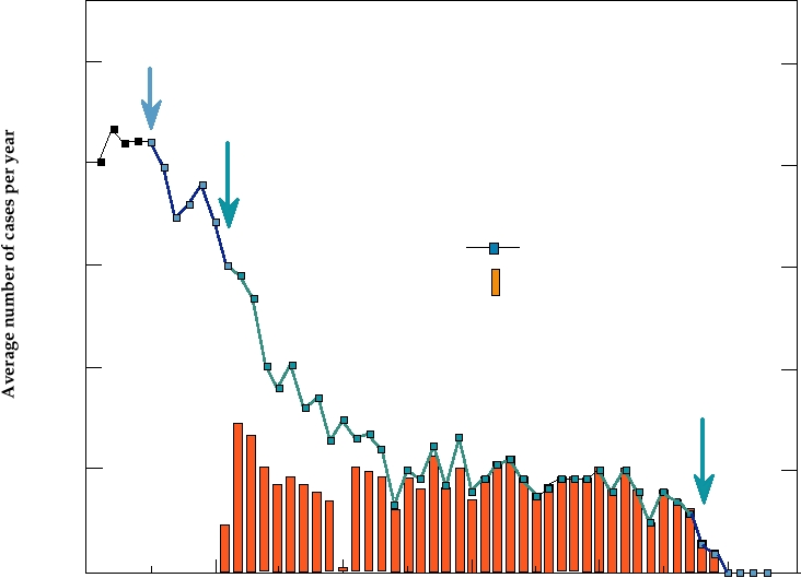
Introduction of the Salk and Sabin vaccines in the 1950s
susceptible but very young children is less likely to cause
and 1960s led to the elimination of poliovirus in the United
poliomyelitis. Statistics of the fraction of young children
States over a period of about 2 decades (Fig. 3.4) and more
who contract poliomyelitis in societies in which the virus
recently has led to the elimination of poliovirus throughout
is endemic, rather than epidemic, are not well defined, in
the Americas. The Salk vaccine, which was the first to be
part because of the high death rate of children in such socie-
developed, is an inactivated virus vaccine that is given as a
ties due to many infectious diseases. However, surveys con-
series of injections. Introduction of this vaccine resulted in a
ducted in the twentieth century of lameness in populations,
rapid decrease in the number of poliovirus cases. However,
most of which is probably due to paralytic polio, found simi-
because the vaccine induces circulating antibodies but little
lar extents of lameness whether the virus was endemic or
in the way of mucosal immunity (see Chapter 10), it prevents
epidemic.
poliomyelitis, the disease, by preventing spread of the virus
In any event, it is clear that changes in human behavior
from the gastrointestinal (GI) tract to the CNS, but not infec-
can bring about serious complications relating to infectious
tion of the GI tract by the virus. The virus thus remained in
disease, and such scenarios have recurred many times during
circulation. The Sabin vaccine, introduced shortly thereafter,
the last century. However, it is important to note that although
Cases of Poliomyelitis in the United States from 1951-2004
Salk Vaccine
(IPV)
100,000
Sabin Vaccine
(OPV)
10,000
Number of cases/year
1000
Vaccine-related cases
100
Sabin Vaccine
Discontinued
10
1950
1960
1970
1980
1990
2000
Year
FIGURE 3.4 Total number of cases of poliomyelitis in the United States from 1951 to 2004 and the number of vaccine-
related cases after the introduction of the live virus Sabin vaccine. IPV is inactivated polio vaccine; OPV is oral polio
vaccine. Data from N. Nathanson et al. (1996) p. 556 and from Morbidity and Mortality Weekly Report (MMWR). Note that
the scale is logarithmic, with each division portraying 10 times as many cases as the one below.
is a live attenuated vaccine that is given orally. Attenuation
(oral administration of relatively small doses of live virus
was achieved by blind passage of the virus followed by test-
versus injection of large amounts of inactivated virulent
ing of the resulting virus in monkeys. The changes resulting
virus), making it suitable for widespread use in developing
from passage are now known and are shown in Table 3.4;
countries. Worldwide use of this vaccine has resulted in the
two changes are sufficient to make the virus avirulent in the
eradication of wild-type poliovirus in the United States and
case of types 2 and 3. The introduction of the Sabin vaccine
throughout the Americas (the last case of indigenous polio-
led to a further rapid decline in paralytic poliomyelitis. This
virus infection in the Americas occurred in Peru in 1991).
vaccine has the drawback that it induces a very small number
Poliovirus is in the process of being eradicated in other parts
of cases of paralytic disease, termed vaccine associated para-
of the world, although it is still endemic in areas of Africa
lytic poliomyelitis (VAPP), that result from reversion of the
and Asia. With the extirpation of polio in the United States,
attenuated virus to virulence. The incidence rate is about 1
the use of Sabin vaccine was discontinued in this country
per million persons inoculated, and there were about 10 such
and it has been replaced with the Salk vaccine, in order to
cases per year in the United States until use of this vaccine
eliminate VAPP.
was discontinued in the year 2000 (Fig. 3.4). The efficacy of
As described before, polioviruses undergo recombina-
the Sabin vaccine is very high, however, because it induces
tion with other enteroviruses. It is important, therefore, to
mucosal immunity as well as other forms of immunity. It
note that the mutations in the Sabin vaccines that render the
prevents subsequent infection by the wild-type virus, thus
virus attenuated are all found in the structural region of the
allowing eradication of the wild-type virus if coverage is
genome (Fig. 3.6). Thus, recombination with other entero-
sufficiently broad. In addition, it is much cheaper and sim-
viruses cannot restore the virulence of the virus. Reversion
pler to manufacture and administer than the Salk vaccine
to virulence requires the back mutation of the attenuating
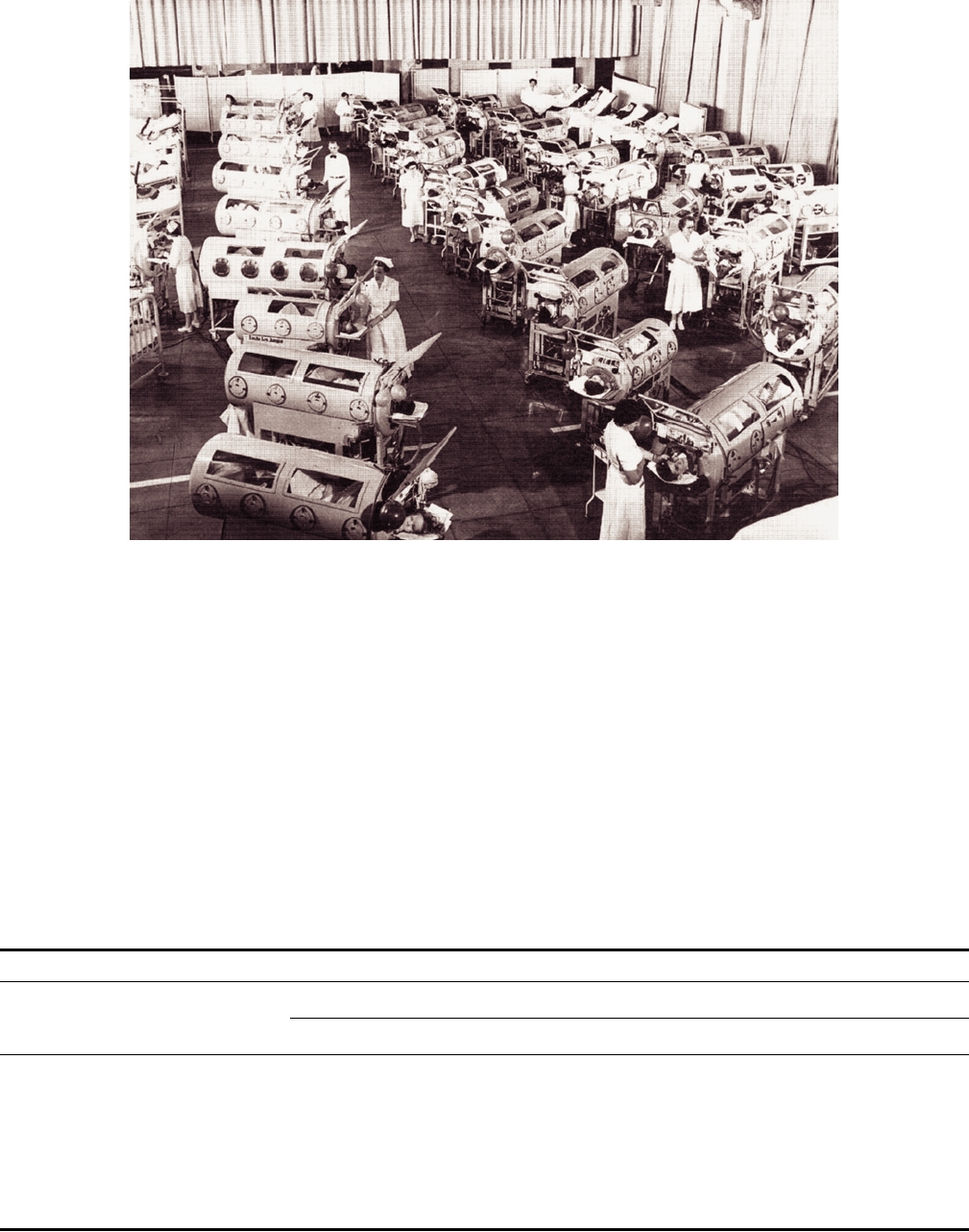
FIGURE 3.5 Ward of iron lungs and rocking beds at the poliomyelitis rehabilitation center in Rancho Los Amigos,
California. From Halstead (1998) with the permission of the author and the publisher.
mutations in the vaccine viruses or, conceivably, recombina-
and the current difficulties in complete eradication of polio
tion between two attenuated poliovirus strains that eliminates
have important lessons for us. The original Salk vaccine was
the attenuating mutations. In the latter case, however, it is
incompletely inactivated because the science of virology
unlikely that most recombinants would be virulent because
was insufficiently developed to assay for minute amounts of
of the incompatibility of the various nonstructural proteins
residual live virus in solutions containing very high concen-
with one another.
trations of virus. The result was that this vaccine caused a
small number of cases of poliomyelitis, but in retrospect the
Development of the Polio Vaccine
riskreward ratio was favorable because of the significant
The polio vaccine has been enormously successful in con-
decline in natural infections (Fig. 3.4). As soon as the prob-
trolling this virus scourge, but the history of its development
lem was recognized, more stringent methods of inactivation
TABLE 3.4
Characteristics of Poliovirus Vaccines
Salk vaccine
Inactivated wild-type poliovirus (three types)
Sabin vaccine
Live poliovirus, attenuated by mutations in:
Type 1
Type 2
Type 3
nt
aa
nt
aa
nt
aa
5′NTR
A480G
--
G481A
--
C472U
--
VP1
G2795A
A106T
C2909U
T143I
U2 493C
I6T
C2879U
L134F
VP3
U2438A
L225M
C2034U
S91F
VP4
G935U
A65S
P1
P2
P3
5 NTR
3B
3D
AAAn
VP2
VP3
4
2A
2B
2C
3A
3C
VP1
A106T
SABIN 1 A65S
L225M
L134F
U2438A C2879U
A480G G935U
G2795A
T143I
SABIN 2
G481A
C2909U
I6T
SABIN 3
S91F
C472U
C2034U
U2493C
0
1
2
3
4
5
6
7
kb
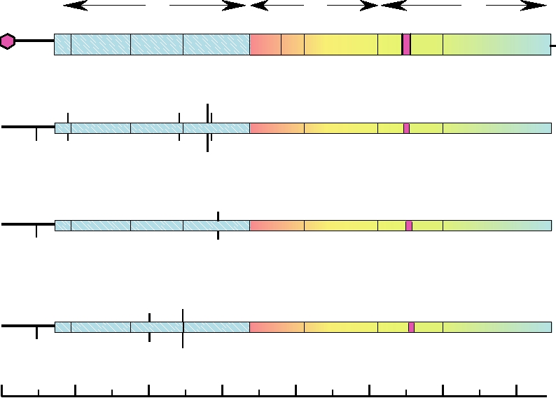
FIGURE 3.6 Diagrams of the genomes of the Sabin vaccine strains of poliovirus types 1, 2, and 3 showing the locations
of the attenuating mutations.
were quickly developed that resulted in complete inac-
Health Organization (WHO) initiated a campaign, the
tivation of the infectivity of the virus, solving this prob-
Global Polio Eradication Initiative, to eradicate poliovi-
lem and serving as an example for development of other
rus worldwide by the year 2000. Although falling short of
vaccines. The introduction of the Salk vaccine, although
this goal, significant progress has been made. The number
enormously successful in controlling polio, also suffered
of polio cases worldwide fell from an estimated 300,000+
from early problems when it was found that early lots were
cases in the mid 1980s to fewer than 3000 by 2000 and sub-
contaminated with the monkey virus SV40 (described in
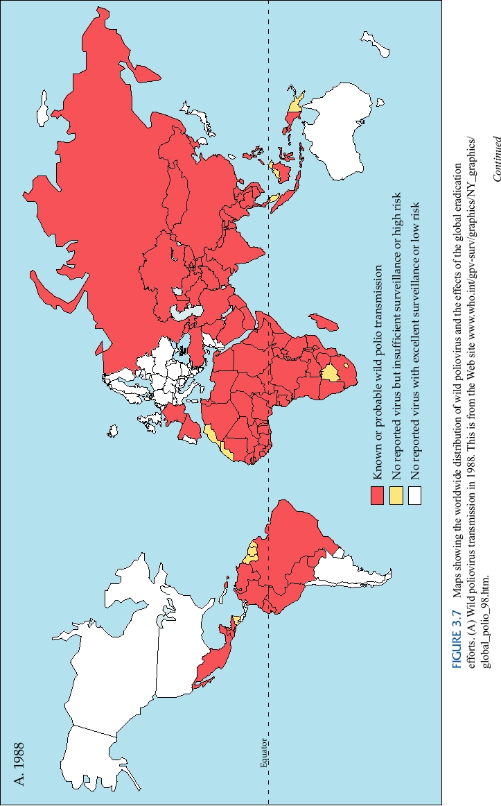
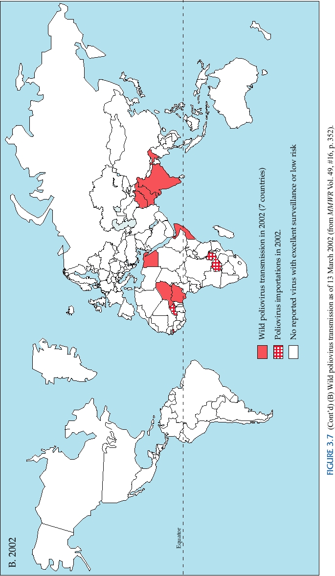
sequently to still lower levels (Fig. 3.7). In 2000 the Centers
Chapter 7). Infection of humans by this virus appears to be
for Disease Control and Prevention (CDC) said that 2971
benign, although there is some evidence that, very rarely,
cases were reported of which 719 were confirmed by labo-
brain tumors may be associated with infection. In any event,
ratory analysis. In 2001, 537 cases were reported of which
this episode brought to light the issue of adventitious con-
473 were confirmed by laboratory analysis, and these cases
tamination of cell cultures with viruses that infect the host
occurred in just 10 countries. It appeared that eradication
supplying tissues for culture. A third problem that arose
would be achieved soon. The eradication campaign hit a
during development of polio vaccines was the infection of
snag recently, however, when Muslim clerics in Nigeria
a number of laboratory workers in Germany with Marburg
claimed that the vaccine could cause AIDS or infertility.
virus. These workers were employed in the isolation of
In 2003, officials in some parts of Nigeria suspended local
cells from the kidneys of wild-caught monkeys that were
vaccination programs, and an epidemic of poliomyelitis in
to be used in propagating polioviruses, and some of the
Nigeria resulted that then spread to neighboring countries
monkeys were infected with Marburg virus, at that time an
that had been free of polio (Fig. 3.8). By 2005 polio had
unknown virus. Several people died in the ensuing epidemic
spread to a total of 16 countries that had previously been
(described in Chapter 4).
polio free. Further setbacks in the polio vaccination ini-
tiative have resulted from civil unrest in Sudan and other
Eradication of Polioviruses
countries that resulted in interference with vaccine cam-
Introduction of the Sabin vaccine led to the eradication
paigns and the reestablishment of poliovirus transmission.
of poliovirus from the Americas, and in 1988 the World
Health ministers from Africa are stepping up vaccination
Tunisia
Morocco
Algeria
Western Sahara
Libya
Egypt
Mauritania
Mali
Niger
Chad
Sudan
Senegal
Gambia
Burkina Faso
Guinea Bissau
Guinea
Nigeria
Ethiopia
Sierra Leone
Ivory Coast
Ghana
Liberia
Cameroon
Somalia
Benin
Togo
Equatorial
Uganda
Guinea
Kenya
Congo
Gabon
Rwanda
Zaire
Burundi
Cabinda
Tanzania
Countries with cases of poliomyelitis
Malawi
Angola
2002
Zambia
Mozambique
2003
Zimbabwe
Caprivi Strip
2004
Namibia
Botswana
Swaziland
South Africa
Lesotho
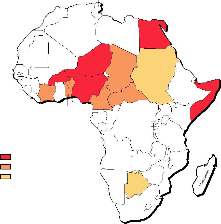
FIGURE 3.8 Reemergence of poliomyelitis in Africa 20022004. Data from MMWR.
programs to reestablish control of poliovirus transmission,
in nonimmunized or incompletely immunized contacts
but these situations illustrate problems that result from the
(e.g., 21 cases in Hispaniola in 20002001), illustrating the
continuing conflicts among societies.
potential for continued outbreaks arising from vaccination.
The Salk vaccine could still be used in developed countries,
Should Routine Poliovirus Immunization Be Eliminated?
but it seems unlikely that routine administration of Salk
If poliovirus is finally eradicated worldwide, should
vaccine would be used in developing countries. It should
vaccination against the virus be scaled back? Limiting vac-
be possible to design new attenuated viruses for vaccine
cination would be important because of the residual viru-
purposes that would be safer than the Sabin vaccine, but
lence of the vaccine virus. Virulent revertants of vaccine
in the absence of poliovirus epidemics no drug company
virus not only cause a small number of individual cases
would want to undertake the very expensive development
(Fig. 3.4), but have led to small epidemics of poliomyeli-
of a new vaccine, especially in view of legal problems that
tis when the virulent virus, derived by reversion, circulates
would be sure to arise.
Associated problems are the difficulties in being sure that
infection of mice, it has been found that the virus can
poliovirus is truly eradicated and the possibility that virulent
persist in neurons in a latent state for at least 12 years.
poliovirus could reemerge. It is known that immunodeficient
There is no evidence that poliovirus might similarly per-
children who have received the vaccine virus can continue
sist in humans for 40 years, however, and such persis-
to secrete virus for long periods of time, during which the
tence seems unlikely. Other possible explanations for the
virus may revert to virulence. Even wild-type poliovirus can
failure of motor neurons in post-polio syndrome have also
circulate silently because most infections are inapparent. In
been suggested. Fortunately, paralytic poliomyelitis and
regions where poliomyelitis has reemerged following inter-
its sequelae may soon be a thing of the past.
ruption of vaccination campaigns, it has been found in some
cases that the virus had been circulating for at least 2 years
before reemergence. In addition, poliomyelitis can be caused
Other Enteroviral Diseases of Humans
by enteroviruses other than poliovirus (see later), a possible
As described, 62 human enteroviruses other than polio-
source of confusion in diagnosis.
viruses are currently recognized in the ICTV catalog and
If vaccination were terminated and poliovirus were to
classified as serotypes of 4 different species. Although most
reemerge in a naïve population, it could lead to a widespread
of these have been known for 50 years, it is only recently
epidemic. Possible sources of reemerging virus include new
that the association of many of these viruses with signifi-
strains that might arise from other enteroviruses, circulating
cant human illness has been shown. In fact, it has now been
strains of wild poliovirus or vaccine-derived virus that have
established that most enteroviruses do cause disease, and
escaped detection, or the inadvertent or deliberate release of
many of them cause significant episodes of serious disease
wild poliovirus by escape from a laboratory or introduction
(Table 3.5). Study of disease caused by these viruses has
by terrorists. It would be necessary to maintain stockpiles
been complicated by the fact that there are so many entero-
of polio vaccine to counter such threats, and in the case of
viruses, of which at least some have multiple strains that
the Salk vaccine this stockpiling itself could serve as a pos-
may differ in disease-causing potential, and by the fact that
sible source of accidental escape of the wild virus. Since
serious disease is an uncommon complication of infection
the events of 9/11, it is also obvious that terrorists would
by most enteroviruses (even for poliovirus most infections
have no compunctions about releasing a virulent virus into
do not result in significant disease). This has made it dif-
the U.S. population if such a virus could be obtained. Thus,
ficult to ascribe any particular disease to infection by any
even if (when?) poliovirus is eradicated, the policies with
particular virus. However, even though serious disease is
respect to vaccination will require careful consideration.
an uncommon complication, enteroviral infections are very
Post-Polio Syndrome
common, and the total number of cases of disease caused by
these viruses is large. These illnesses include very infrequent
Although poliovirus has been eradicated from devel-
paralytic disease essentially indistinguishable clinically
oped countries, there is a large cohort of people infected
from that caused by poliovirus; myocarditis and pericarditis
in the 1950s who are or were paralyzed. Many paralyzed
(caused especially by the Coxsackie B viruses) that is usu-
poliomyelitis patients were ultimately able to resume
ally subclinical but can be acute and result in significant car-
almost normal activities. Through a process of axonal
diac compromise; aseptic meningitis; encephalitis; hepatitis;
sprouting and reenervation of muscles by the motor neu-
the common cold (perhaps a quarter of summer colds are due
rons that survived the infection, many learned to walk and
to enteroviruses); diarrheal disease; febrile illnesses; rash;
use their previously paralyzed limbs. In many, recovery
hand-foot-and-mouth disease (a common childhood illness
was effectively complete. However, a syndrome called
caused by several serotypes in human enterovirus A); and
post-polio syndrome has emerged to plague a significant
epidemic acute hemorrhagic conjunctivitis (an epidemic
fraction, perhaps 40%, of the survivors of paralytic polio-
disease caused by enterovirus 70 that appeared recently and
myelitis. This syndrome appears 3040 years after polio
spread around the world). The Coxsackie B viruses are also
infection and is characterized by fatigue, pain, and weak-
associated epidemiologically with juvenile onset diabetes in
ness. The weakness may be severe enough to require the
humans but how (or even whether) they cause diabetes is still
use of a wheelchair. The syndrome results from the degen-
unresolved. There are no vaccines for any of these viruses.
eration of motor neurons, but the reasons for the degenera-
tion are not clear. The favored hypothesis is that it is the
result of overuse of the surviving motor neurons, which
are forced to do the work of many. A second possibility is
Genus Rhinovirus
that the surviving neurons were damaged by the original
poliovirus infection and fail prematurely. A third, albeit
The human rhinoviruses are the causative agents of about
unlikely, possibility is that poliovirus persists in neu-
half of human colds, the most characteristic symptom of
rons and is somehow reactivated, even in the presence of
which is rhinitis (inflammation of the nasal mucous mem-
anti-polio antibody. In model studies using Sindbis virus
brane and characterized by a runny nose). Other viruses that
TABLE 3.5
Clinical Syndromes Associated with Human Enteroviruses
Clinical syndrome
Poliovirus
Enterovirus A
Enterovirus B
Enterovirus C
Enterovirus D
Paralysis
Types 1, 2, 3
Coxsackie A7, A9
Coxsackie B2B5
--
Enterovirus 70
Enterovirus 71
Echoviruses 4, 6, 9, 11, 30
Aseptic meningitis
--
Coxsackie A2, A4, A7,
Coxsackie B1B6
--
--
A9, A10
All echoviruses except
12, 24, 26, 29, 32, 33
Pericarditis, myocarditis
--
--
Coxsackie B1B5
--
--
Echoviruses 1, 6, 9, 19
Encephalitis
--
Enterovirus 71
Coxsackie B1B5
--
Enterovirus 70
Echoviruses 2, 6, 9, 19
Hepatitis
--
Coxsackie A4
Coxsackie A9, B5
--
--
Echovirus 4, 9
Upper respiratory
--
--
Coxsackie B4, B5
Coxsackie A21, A24
Enterovirus 68
disease, pneumonia
Hand, foot, and
--
Enterovirus 71
--
--
--
mouth disease
Coxsackie A5, A10, A16
Acute hemorrhagic
--
--
--
Coxsackie A24
Enterovirus 70
conjunctivitis
Undifferentiated
Types 1, 2, 3
--
Coxsackie B1B6
--
--
febrile illness
serve as major causes of the common cold include some
years have not been done to establish whether immunity
of the enteroviruses, just described, and the coronavi-
to a particular rhinovirus following infection is long lived.
ruses, described later. One hundred serotypes of human
For the same reasons, there are no vaccines for any of these
rhinoviruses are currently recognized. Eighteen of these
viruses.
have been assigned to the species human rhinovirus A
Rhinoviruses replicate in the upper respiratory tract and
and three of them to human rhinovirus B. The remain-
are transmitted by direct person-to-person contact. Coughing
ing 79 serotypes have not yet been assigned to a spe-
and sneezing, common syndromes of rhinovirus infection,
cies. There are also three serotypes of bovine rhinovirus
help spread the virus to nearby contacts. It is not clear how
known to exist, and there are rhinoviruses for other ani-
much of the spread is due to aerosolization of the virus on
mals that have as yet to be well characterized. In general,
coughing or sneezing followed by inhalation of the aero-
rhinoviruses are specific for a particular species or for
solized virus by a susceptible contact, and how much is due
a limited range of species, and this restriction appears
to contact with mucus that contains virus, such as by hand-
to work at the level of receptors required for virus entry
shake or contact with contaminated doorknobs, followed by
(see Chapter 1).
transmission of the virus to mucosal membranes in the nose
The 100 serotypes of human rhinoviruses are not cross
or the mouth.
protective and the result is that we are subject to many
It is an interesting and informative historical fact that
rhinovirus colds during our lifetimes. Young children, not
early attempts to isolate rhinoviruses using standard cell cul-
having been exposed to rhinoviruses and other viruses
ture techniques were unsuccessful. Most cells in the body
that cause colds, contract many colds a year. Adults, hav-
are maintained at 37°C at a pH of 7.4, and cells in culture are
ing become immune to many of these viruses through
normally maintained under these conditions. However, cells
hard experience, have fewer colds per year, usually only
in the upper respiratory tract are maintained at a lower tem-
about one. However, the extent and duration of immunity
perature, about 33°C, because the inhalation of outside air
to a particular rhinovirus induced by infection are not well
through the upper respiratory tract keeps this area cool, and
established. There are so many rhinoviruses (and although
at a pH significantly less than 7.4 because of the high con-
rhinoviral disease may be miserable it is not life threaten-
centration of CO2 in expired air. Rhinoviruses replicate well
ing) that detailed studies on cohorts of people over many
in cultured cells under these altered conditions and appear
to require the lower temperature and lower pH for efficient
the viruses are unrelated. For reference, Table 3.6 presents
growth. In part because of this, rhinovirus infection is limited
a description of the currently known viruses whose name
to the upper respiratory tract, and rhinoviruses almost never
includes hepatitis. Figure 3.9 shows the incidence of hepati-
cause lower respiratory tract infections.
tis in the United States in 1997 caused by hepatitis viruses A,
It is also of interest that rhinoviruses are sensitive to very
B, and C, which are the most important causes of viral hepa-
low pH, and infectivity is destroyed by exposure to pH 3.
titis in the United States. Identification of which hepatitis
The related polioviruses, however, survive exposure to pH
virus is responsible for any specific case of hepatitis requires
2, which is necessary because, being enteroviruses, they
immunologic tests or virus isolation, because symptoms are
must survive passage through the stomach in order to infect
similar.
an animal.
Hepatitis A virus is a picornavirus and will be considered
here. The other viruses will be considered when their respec-
tive families are introduced.
Genus Cardiovirus
The Cardiovirus genus consists of several viruses of mice
Hepatitis A Virus
of which encephalomyocarditis virus (EMC) has been exten-
Hepatitis A virus (HAV) is a causative agent of infectious
sively studied as a model picornavirus. It is closely related to
hepatitis in humans. The virus is worldwide in distribution.
other picornaviruses (Fig. 3.1) although differing in certain
Only one serotype is known, but isolates from different areas
important characteristics. The EMC IRES has proved more
or different times can be grouped into different genotypes or
useful than the poliovirus IRES in experiments that require
strains. The most distantly related HAV isolates share about
polycistronic mRNAs or that express proteins in a cap-inde-
75% nucleotide sequence identity, but most isolates are
pendent fashion in vertebrate expression systems. Theiler's
much more closely related. HAV is a typical picornavirus
virus, another member of this genus, causes demyelinating
but is an outlier in the family (Fig. 3.1). It shares only 28%
disease in mice and has been extensively studied as a model
amino acid identity in its structural proteins with any other
for multiple sclerosis in humans.
picornavirus, whereas most picornaviruses are more closely
related to one another.
Genus Hepatovirus
The number of cases of hepatitis A in the world has been
estimated to be more than 1.4 million each year. In 1998
Hepatitis in Humans
in the United States, for example, ∼37,000 cases of hepa-
Many different viruses, belonging to several virus fami-
titis were reported, of which two-thirds were diagnosed as
lies, are known to cause hepatitis (inflammation of the liver)
caused by HAV. HAV is spread through contaminated food
in humans. These different viruses have different modes
and water. Filter-feeding shellfish like oysters are known to
of transmission and cause illness of different degrees of
concentrate the virus, and consumption of raw shellfish has
severity (although all hepatitis is serious) that results from
been the cause of many epidemics of hepatitis A. A 2003
destruction of liver cells caused by growth of these viruses
epidemic of more that 500 cases in Pennsylvania was caused
in the liver as a target organ. Hepatitis is characterized by
by consumption of green onions that are believed to have
fatigue and other symptoms that result from inadequate liver
been contaminated during harvest. Infection by HAV usually
function, and it may be fatal if sufficient destruction of the
results in a self-limited illness in which the patient recovers
liver takes place. A characteristic feature of acute hepatitis is
with relatively few sequelae. The illness can be quite serious,
the presence of elevated levels of liver enzymes circulating
even fatal, however, because 90% of the liver tissue can be
in the blood that results from the destruction of liver cells.
destroyed by virus infection, and liver function is severely
Many cases of hepatitis are accompanied by jaundice (turn-
impaired until the liver recovers. The seriousness of disease
ing yellow) because of the destruction of the liver, which is
is age dependent. Very young children suffer little disease
responsible for clearing bilirubin from the blood.
but with advancing age infection by the virus becomes more
The viruses whose primary disease syndrome in humans
serious. The mortality rate in children younger than 14 is
is hepatitis, and which therefore target the liver as the princi-
only 0.1%, but HAV infection in people older than 40 results
pal or only organ infected, or viruses that are closely related
in a fatality rate of 2.1%.
to viruses that cause such hepatitis, have historically been
Before the introduction of a vaccine, the only prophylaxis
named hepatitis virus followed by a letter, in the order of
for HAV was injection of immune gamma globulin, which
isolation. Thus we have hepatitis A virus, the first to be iso-
provided protection from the virus for a few weeks. Two
lated, hepatitis B virus, the second, and so forth. Because
inactivated virus vaccines against HAV were licensed in the
these viruses belong to a number of different families, con-
mid 1990s that have been found to give long-lived protec-
fusion can arise because of the similar names even though
tion. Results from a clinical trial in Thailand that showed the
TABLE 3.6 Causative Agents of Viral Hepatitis in Humans
Annuala U.S. acute
Chronic
Family/
Genome type/
Long-term
cases/deaths
hepatitis (millions
Virus
genus
size in kb
Transmission
Chronicity?
effects
in 2004
of cases) U.S. / World
Hepatitis A
Picornaviridae/
ss (+) RNA/
Fecaloral
Very little
Few if any
5683/76
0/0
Hepatovirus
7.5 kb
ds DNA (RT)/b
HCCc, cirrhosis
Hepatitis B
Hepadnaviridae/
Parenteral, sexual,
10% of adults,
6212/659
1.2/300400
Orthohepadno-
3.2 kb
vertical
90% of
virus
neonates
HCCc, cirrhosis
Hepatitis C
Flaviviridae/
ss (+) RNA/
Parenteral, sexual,
>50%
720/4321
3/50100
Hepacivirus
9.4 kb
vertical
Hepatitis D
Deltavirus
ss, circular
Parenteral, (sexual,
Yes
Exacerbates
7500/
0.07/?
RNA/1.7 kb
vertical?)
symptoms
1000
of Hep B
Hepatitis E
Hepeviridae/
ss (+) RNA/
Fecaloral
No
Few if any
Very rare
0/0
Hepevirus
7.5 kb
Hepatitis Fd
?
Hepatitis G
Flaviviridae/
ss (+) RNA/
Parenteral, other?
Yes
??
??/none
??
Hepacivirus
9.4 kb
a
Data from MMWR Summary of Notifiable Diseases--2004. It is noteworthy that acute cases of both hepatitis A and hepatitis B have declined significantly
since 1990 with the introduction of vaccines that are now in widespread use; see Figure 3.9 below.
b
RT is reverse transcriptase. Nucleic acid in virion is partially ds DNA, consisting of a full-length minus-strand DNA of 3.2 kb, and an incomplete
plus-strand DNA that is variable in length.
c
HCC, Hepatocellular carcinoma.
d
Isolate from a fulminant case of hepatitis, not further characterized.
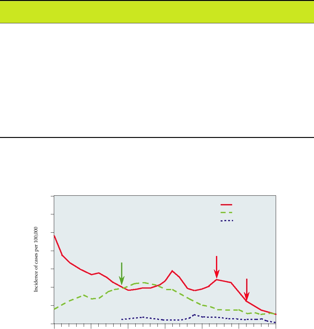
35
Hepatitis A
Hepatitis B
30
Hepatitis C
25
20
Hep A vaccine
Hep B vaccine
15
ACIP guidelines
10
5
0
1973
1978
1983
1988
1993
1998
2003
Year
FIGURE 3.9 Incidence (cases per 100,000 population) of viral hepatitis in the United States between 1973 and 2003.
Hepatitis A incidence was the lowest ever in 2004, but there has been a trend for cyclic increases every decade, and future
increases could occur. However, with the expansion of recommended vaccination to include children in all communities
where the incidence was consistently above the national average (1999 ACIP guidelines), the incidence of HAV has
continued to plummet. No vaccine for hepatitis C exists, but an antibody test for hepatitis C was first introduced in May
1990. This graph is adapted from MMWR Vol. 52, #54, Summary of Notifiable Diseases United States 2003.
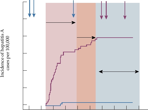
12
3
12
3
Dose
300
Start
250
Surveillance
Booster
Surveillance
200
Control Group
150
Controlled Trial Ends
100
50
Hep A Vaccine Group
0
0
200
400
600
800
Time after dose 1 in days
FIGURE 3.10 Controlled trial of a hepatitis A vaccine in Thailand. Children were divided into two groups, and at the
times indicated by the blue arrows the vaccine group was given hepatitis A vaccine and the control group was given hepatitis
B vaccine. There were two periods of surveillance for cases of hepatitis A, indicated by the pink overlays. The controlled
trial ended at 540 days when the control group was given hepatitis A vaccine at the times indicated by the magenta arrows
and the vaccine group was given hepatitis B vaccine. Adapted from Figure 3 in Innis et al. (1994).
efficacy of this vaccine are shown in Fig. 3.10, as an exam-
interesting differences in the replication of this virus from
ple of the type of data that can be obtained in clinical trials.
those of other picornaviruses, such as the presence of VP0
The introduction of these vaccines and the recommendation
in virus particles rather than the cleaved products VP2 and
in 1996 of routine childhood vaccination has resulted in a
VP4 (see Fig. 3.2). The worldwide burden of human gas-
steady decline in the rate of hepatitis A in the United States.
troenteritis caused by Aichi virus or closely related viruses
In 2003 there were 7653 cases reported (2.7 per 100,000
is unknown at present. As described later, many viruses
population), and childhood vaccination appears to have been
belonging to a number of virus families cause epidemic gas-
important in the control of this disease. Of interest is the
troenteritis in humans, and sorting out the causative agents is
finding that hepatitis A rates were much higher in the west-
difficult and requires time.
ern United States before the introduction of the vaccines, but
A virus related to Aichi virus appears to be widespread
the rates are now similar across the United States, and an
in cattle in the Aichi area of Japan. This virus, classified for
increasing proportion of cases occur in adults.
now as a member of the Kobuvirus genus, apparently causes
inapparent infection in cattle and has been called bovine
kobuvirus. Thus, it is possible that kobuviruses are widely
Genus Kobuvirus
distributed in the world and infect a number of species.
In Aichi, Japan in 1989, a stool specimen from a patient
suffering from gastroenteritis associated with consumption
Genus Aphthovirus
of raw oysters was found to contain a new picornavirus, sub-
sequently called Aichi virus. Since then the virus has been
Foot-and-mouth disease viruses (FMDV) belong to
found not only in Japan but also in Pakistani children with gas-
seven currently recognized serotypes. They cause a debili-
troenteritis and in Japanese travelers returning from Southeast
tating disease, foot-and-mouth disease, in cattle and other
Asia with gastroenteritis, and the virus thus appears to be
animals, and are economically important pathogens. FMDV
widespread and to be one of the many viruses that are causa-
was eliminated from the United States many years ago by
tive agents of human gastroenteritis. The virus is sufficiently
the simple expedient of killing all infected animals until
distinct in its sequence from other picornaviruses to be clas-
such time as the virus was extirpated. The last epidemic
sified in a separate genus, Kobuvirus (Fig. 3.1). The name
in the United States occurred in 1929. The virus still cir-
is derived from the Japanese word for bump, because the
culates in Europe, South America, and other parts of the
virion appears bumpy in the electron microscope. There are
world, and the U.S. Department of Agriculture maintains
Search WWH :